Wednesday, July 6, 2011

Cervical Curve and Its Importance
A New Cervical Lordotic Home Traction Device: The Denneroll—An Initial Case Series
Sunday, February 14, 2010 at 6:34AM
CBP Seminars
CBP Seminars

Deed E. Harrison, DC
President CBP Seminars, Inc.
Vice President CBP Non-Profit, Inc.
ICA Nevada State Assembly Rep
Chair PCCRP Guidelines
Editor—AJCC
In a previous issue of the AJCC (2008), I presented the evidence for in office CBP® Technique structural rehabilitative procedures. According to the CBP publications, mirror image® exercises and traction procedures should be performed in-office at least 3-4 times per week for 9-12 weeks in order to obtain significant improvement in abnormal alignment of the spine and improvements in a patient’s chronic disorder(s).1-5
However, the evidence based practice guideline/protocol of care for CBP technique, recommends home exercise and home traction for patients as a supplementary procedure to the in-office intervention program.4,5Also, home traction and exercise may be the only viable means of rehabilitation for patient’s whom live many miles away from a given practice or for schedules that are simply not conducive to regular care.
Problematically, to my knowledge, there exist no formal publications documenting the potential benefit/effect of different types of home traction procedures aimed at rehabilitation of the sagittal plane curvatures of the spine. Thus, the purpose of the present article is to present preliminary information on the immediate effect of one traction session using a new cervical orthotic device termed the Denneroll.
Materials and Methods
The Denneroll (Designed by Adrian Dennewald, D. C. of Australia) is a new cervical sagittal plane orthotic device designed to passively stretch the cervical lordosis into a more lordotic position. See Figure 1. Its unique design allows it to support the upper thoracic
curvature while simultaneously create a 3-point bending extension load on the cervical spine.
Most commonly, the Denneroll has 3 primary positions that are used for aiding in the rehabilitation of the cervical lordosis:
1) The apex of the Denneroll orthotic is placed in the upper cervical region (C2-C4) region. This position allows extension bending of the upper cervical segments while causing slight anterior head translation. An upper neck setup and example x-ray subluxation is shown in Figure 1. The red line represents the ideal curvature after Harrison et al.6 while the black line shows the patient’s subluxated alignment.
2) The apex of the Denneroll orthotic is placed in the mid-cervical region (C4-C6) region. This position allows extension bending of the mid-upper cervical segments while creating a slight posterior head translation. SeeFigure 1.
3) The apex of the Denneroll orthotic is placed in the upper thoracic or lower-cervical region (C6-T1) region. This position allows extension bending of the majority of cervical segments while creating a significant posterior head translation. See Figure 1.
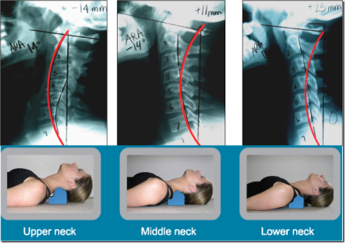
Figure 1. On the top row from left to right, 3 uniquely different subluxated lateral cervical curvatures are shown; the red line represents the ideal curvature of the neck after Harrison et al.1 On the bottom row, three primary placements of the Denneroll cervical orthotic are shown. The Denneroll placement should match both the shape of the cervical curve and the amount of sagittal head translation correction that is desired.
For a preliminary investigation, 11 Chiropractors volunteered for an initial neutral lateral cervical radiograph and completed a neck disability index. One out of the 11 lateral cervical came out with digital artifacts that could not be corrected and was discarded; while another had a normal cervical lordosis on the initial lateral x-ray. This left 9 subjects.
The 9 subjects were asked to lie supine on the floor over the Denneroll orthotic device for 10-13 minutes. Only 1 traction-session was used. The Denneroll location was selected by a trained practitioner.
Following the 10-13 minute traction session, the subjects were asked to relax comfortably for 3-5 minutes without stretching or bending the neck. Once the 3-5 minute interval elapsed, a second neutral lateral cervical radiograph was obtained.
Results
The initial and follow-up lateral cervical radiographs were analyzed with the PostureRay x-ray digitization system. Only 2 of the many reported variables are shown in Table 1. The cervical lordosis using the posterior body margins of C2-C7 and the sagittal plane translation of C2-C7 were recorded. From Table 1 it can be seen that a significant improvement in the cervical lordosis (9.8°) and reduction in sagittal plane head translation (7.4mm) were obtained.
Figure 2 shows subject #9’s initial and after lateral cervical alignment. Here the green semi-circular line represents the ideal cervical lordosis after Harrison et al.6; while the red line represents his lateral cervical alignment.
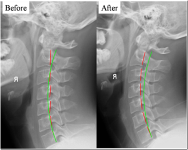
Figure 2. Before and After Denneroll x-rays. With the subject supine, the Denneroll was placed in the lower neck as in Figure 1 for 11 minutes. The following up x-ray was taken after 5 minutes of recovery (no Denneroll). Good improvement in cervical lordosis was found after 1 session indicating likely benefit.
Discussion
It is significant that following only one 10-13 minute session on the Denneroll orthotic device, a significant improvement in both the cervical lordosis and anterior head translation. Obviously the results presented herein are preliminary and follow-up should be and will be performed on this device.
From Table 1, the astute reader will see that a couple of the subjects showed remarkable change in lordosis while a couple of subjects showed little only slight change. This type of situation is typical of any/all treatment devices and is due to many variables:
· The elasticity of the individual subject’s tissues,
· The age of the subject,
· The state of degenerative joint disease-stiffness of the tissues,
· The shape of the thoracic curvature,
· Improper application during traction,
· Perhaps the device just won’t work for some individuals, etc.
Most of the above variables can be overcome with continued effort on the patient’s and the doctor’s part. However, as with all interventions, there is no such thing as a one size fits all.
To me, the information presented herein, is preliminary data indicating the Denneroll orthotic may be a viable home traction device to supplement a CBP Chiropractors in office rehabilitative treatments. When the shape of the cervical curve indicates, the Denneroll could be used on off days from office treatments; and in difficult cases, it could be used daily once tolerance is developed.
Hopefully, the information presented will stimulate further research into the effects of home traction units. After all the majority of us (including me) recommend home products to our patients and believe in their effects; but wouldn’t it be nice to know?
Note: If you would like more information on the Denneroll Cervical Orthotic device contact:drdeed@idealspine.com or see www.idealspine.biz.
References
1. Harrison DD, et al. J Manipulative Physiol Ther 1994;17(7):454-464.
2. Harrison DE, et al. Arch Phys Med Rehab 2002; 83(4): 447-453.
3. Harrison DE, et al. J Manipulative Physiol Ther 2003; 26(3): 139-151.
4. Oakley PA, et al. J Canadian Chiro Assoc 2005; 49(4):270-296.
5. Harrison DE, Harrison DD, Haas JW. CBP® Structural Rehabilitation of the Cervical Spine. CBP Seminars, 2002; pgs:147-151. ISBN 0-9721314-0-X.
6. Harrison DD, et al. Spine 2004; 29:2485-2492.
President CBP Seminars, Inc.
Vice President CBP Non-Profit, Inc.
ICA Nevada State Assembly Rep
Chair PCCRP Guidelines
Editor—AJCC
In a previous issue of the AJCC (2008), I presented the evidence for in office CBP® Technique structural rehabilitative procedures. According to the CBP publications, mirror image® exercises and traction procedures should be performed in-office at least 3-4 times per week for 9-12 weeks in order to obtain significant improvement in abnormal alignment of the spine and improvements in a patient’s chronic disorder(s).1-5
However, the evidence based practice guideline/protocol of care for CBP technique, recommends home exercise and home traction for patients as a supplementary procedure to the in-office intervention program.4,5Also, home traction and exercise may be the only viable means of rehabilitation for patient’s whom live many miles away from a given practice or for schedules that are simply not conducive to regular care.
Problematically, to my knowledge, there exist no formal publications documenting the potential benefit/effect of different types of home traction procedures aimed at rehabilitation of the sagittal plane curvatures of the spine. Thus, the purpose of the present article is to present preliminary information on the immediate effect of one traction session using a new cervical orthotic device termed the Denneroll.
Materials and Methods
The Denneroll (Designed by Adrian Dennewald, D. C. of Australia) is a new cervical sagittal plane orthotic device designed to passively stretch the cervical lordosis into a more lordotic position. See Figure 1. Its unique design allows it to support the upper thoracic
curvature while simultaneously create a 3-point bending extension load on the cervical spine.
Most commonly, the Denneroll has 3 primary positions that are used for aiding in the rehabilitation of the cervical lordosis:
1) The apex of the Denneroll orthotic is placed in the upper cervical region (C2-C4) region. This position allows extension bending of the upper cervical segments while causing slight anterior head translation. An upper neck setup and example x-ray subluxation is shown in Figure 1. The red line represents the ideal curvature after Harrison et al.6 while the black line shows the patient’s subluxated alignment.
2) The apex of the Denneroll orthotic is placed in the mid-cervical region (C4-C6) region. This position allows extension bending of the mid-upper cervical segments while creating a slight posterior head translation. SeeFigure 1.
3) The apex of the Denneroll orthotic is placed in the upper thoracic or lower-cervical region (C6-T1) region. This position allows extension bending of the majority of cervical segments while creating a significant posterior head translation. See Figure 1.

Figure 1. On the top row from left to right, 3 uniquely different subluxated lateral cervical curvatures are shown; the red line represents the ideal curvature of the neck after Harrison et al.1 On the bottom row, three primary placements of the Denneroll cervical orthotic are shown. The Denneroll placement should match both the shape of the cervical curve and the amount of sagittal head translation correction that is desired.
For a preliminary investigation, 11 Chiropractors volunteered for an initial neutral lateral cervical radiograph and completed a neck disability index. One out of the 11 lateral cervical came out with digital artifacts that could not be corrected and was discarded; while another had a normal cervical lordosis on the initial lateral x-ray. This left 9 subjects.
The 9 subjects were asked to lie supine on the floor over the Denneroll orthotic device for 10-13 minutes. Only 1 traction-session was used. The Denneroll location was selected by a trained practitioner.
Following the 10-13 minute traction session, the subjects were asked to relax comfortably for 3-5 minutes without stretching or bending the neck. Once the 3-5 minute interval elapsed, a second neutral lateral cervical radiograph was obtained.
Results
The initial and follow-up lateral cervical radiographs were analyzed with the PostureRay x-ray digitization system. Only 2 of the many reported variables are shown in Table 1. The cervical lordosis using the posterior body margins of C2-C7 and the sagittal plane translation of C2-C7 were recorded. From Table 1 it can be seen that a significant improvement in the cervical lordosis (9.8°) and reduction in sagittal plane head translation (7.4mm) were obtained.
Figure 2 shows subject #9’s initial and after lateral cervical alignment. Here the green semi-circular line represents the ideal cervical lordosis after Harrison et al.6; while the red line represents his lateral cervical alignment.

Figure 2. Before and After Denneroll x-rays. With the subject supine, the Denneroll was placed in the lower neck as in Figure 1 for 11 minutes. The following up x-ray was taken after 5 minutes of recovery (no Denneroll). Good improvement in cervical lordosis was found after 1 session indicating likely benefit.
Discussion
It is significant that following only one 10-13 minute session on the Denneroll orthotic device, a significant improvement in both the cervical lordosis and anterior head translation. Obviously the results presented herein are preliminary and follow-up should be and will be performed on this device.
From Table 1, the astute reader will see that a couple of the subjects showed remarkable change in lordosis while a couple of subjects showed little only slight change. This type of situation is typical of any/all treatment devices and is due to many variables:
· The elasticity of the individual subject’s tissues,
· The age of the subject,
· The state of degenerative joint disease-stiffness of the tissues,
· The shape of the thoracic curvature,
· Improper application during traction,
· Perhaps the device just won’t work for some individuals, etc.
Most of the above variables can be overcome with continued effort on the patient’s and the doctor’s part. However, as with all interventions, there is no such thing as a one size fits all.
To me, the information presented herein, is preliminary data indicating the Denneroll orthotic may be a viable home traction device to supplement a CBP Chiropractors in office rehabilitative treatments. When the shape of the cervical curve indicates, the Denneroll could be used on off days from office treatments; and in difficult cases, it could be used daily once tolerance is developed.
Hopefully, the information presented will stimulate further research into the effects of home traction units. After all the majority of us (including me) recommend home products to our patients and believe in their effects; but wouldn’t it be nice to know?
Note: If you would like more information on the Denneroll Cervical Orthotic device contact:drdeed@idealspine.com or see www.idealspine.biz.
References
1. Harrison DD, et al. J Manipulative Physiol Ther 1994;17(7):454-464.
2. Harrison DE, et al. Arch Phys Med Rehab 2002; 83(4): 447-453.
3. Harrison DE, et al. J Manipulative Physiol Ther 2003; 26(3): 139-151.
4. Oakley PA, et al. J Canadian Chiro Assoc 2005; 49(4):270-296.
5. Harrison DE, Harrison DD, Haas JW. CBP® Structural Rehabilitation of the Cervical Spine. CBP Seminars, 2002; pgs:147-151. ISBN 0-9721314-0-X.
6. Harrison DD, et al. Spine 2004; 29:2485-2492.
Article originally appeared on Chiropractic BioPhysics, American Journal of Clinical Chiropractic (http://www.chiropractic-biophysics.com/).
See website for complete article licensing information.
Friday, May 20, 2011
New Study shows how Chiropractic can Help with Tension Headaches
Forward Head Posture, Cervicogenic Headache, and Anatomical Connection Found Between the Rectus Capitis Posterior Major and the Dura Mater
by Dr. Stephanie Maj
A new study, just published in Spine Journal, reports on an investigation of the muscles of the suboccipital triangle and their relationship to cervicogenic headaches (headaches that are caused by subluxations in the neck.)
This got me thinking of a problem I see in over 75% of patients that come to my office: Forward head posture. This position (see picture) puts the neck in an extended position which puts pressure on the nerves that go to the muscles in the back of neck, right under the skull (the rectus capitus muscle group).
This study reports that those muscles connect to the dura mater. The dura mater is the outermost, toughest, and most fibrous of the three membranes, or meninges, covering the brain and spinal cord.
During the anatomic study of thirteen cadaver specimens, it was discovered that eleven of the 13 specimens had a connection between the rectus capitis posterior major muscle (at C2) and the spinal dura mater. [1] A previous report by Hack (Spine 1995) [2] discussed a connection found between the rectus capitis posterior minor and the dura mater and its relationship to cervicogenic headache.
What is most interesting in this new study is that manual traction of the rectus capitis posterior major resulted in gross movement of the dural sheath from the spinal root level at C2, all the way down to the T1 nerve root. Hack previously suggested that:
“It has been speculated that the function of the muscle dural bridge may be to prevent folding of the dura mater during hyperextension of the neck. Also, clinical evidence suggests that the muscle dural bridge may play an important role the pathogenesis of the cervicogenic headaches.”
The authors of the current study concluded that “various clinical manifestations may be linked to this anatomical relationship.” This is where Chiropractic comes in and the stress put on these upper neck structures from forward head posture becomes an important thing to evaluate.
According to Kapandji (Physiology of the Joints, Volume III), for every inch your head moves forwards, it gains 10 pounds in weight, as far as the muscles in your upper back and neck are concerned. That’s because because they have to work that much harder to keep the head (chin) from crashing onto your chest. This abnormal positioning also forces the suboccipital muscles (the ones that raise the chin) to remain in constant contraction, putting pressure on the 3 suboccipital nerves.
This nerve compression may cause headaches at the base of the skull. Pressure on the suboccipital nerves can also mimic sinus (frontal) headaches. It is these nerves and muscles that have the relationship with the dura mater and therefore the brain and headaches.
If you are suffering from headaches of any kind, take a look at how far your head is in front of your shoulders (they should line up ear over top of shoulder.) Chiropractic has great success correcting this postural abnormality and when corrected, can lead to less pressure on upper neck and significant reduction of headaches.
REFERENCES:
1. Anatomical Connection Between the Rectus Capitis Posterior Major and the Dura Mater
Spine (Phila Pa 1976). 2011 (Jan 27)
2. Anatomic Relation Between the Rectus Capitis Posterior Minor Muscle and the Dura Mater
Spine (Phila Pa 1976). 2011 (Jan 27)
2. Anatomic Relation Between the Rectus Capitis Posterior Minor Muscle and the Dura Mater
Spine 1995 (Dec); 20 (23): 2484-2486
Dr. Stephanie Maj has a thriving family practice in the heart of Chicago. Her clinic is located at 1442 W. Belmont Ave., 1E, Chicago, IL 60657. 773.528.8485. www.communitychiropractic.net

 Posted in Bio-mechanics/Ergonomics, Brain Health, Chiropractic Superiority |
  Tags: Brain Health, Chicago, Chiropractic, Dr. Stephanie Maj, Ergonomics, Headache, Health, Neck Pain
3 Responses to “New Study shows how Chiropractic can Help with Tension Headaches”
- This post was mentioned on Twitter by drbsmarinsky, chirosportmonza. chirosportmonza said: New Study shows how Chiropractic can Help with Tension Headaches – http://goo.gl/XW67W via @Shareaholic
- [...] New Study shows how Chiropractic can Help with Tension Headaches [...]
- [...] more: New study shows how chiropractic can help with tension headache Did you like this? Share it:Tweet This entry was posted in Chiropractic, Chronic pain, Frederick, [...]
Tuesday, May 17, 2011
Why Grains Are Not The Best For Us
As you probably know, we’ve been eating grains, refined or whole, only since the beginning of agriculture some 10, 000 years ago. The problem is that our genes where formed about 2 million years ago and haven’t changed much since then, nor did our digestive systems. Eating structurally and chemically different foods is therefore more often than not asking for problems. Grains are miles away from what our bodies should be processing.
In fact, grains aren’t really a good thing for any mammal. We’re better off leaving them to the birds, who have a system adapted to them.
The problem is that our food system is so skewed in the wrong direction that everybody now thinks that grains, especially whole grains, are healthy and nutritious. Grains, a food group that we didn’t eat for 97% of our human existence are now at the bottom of the USDA food pyramid with a recommended 6 to 11 servings per day. This is amazingly wrong!
Be aware that the reason why governments pushed grains in the first place where economical. They are cheap to produce (although not without environmental costs), they can be stored for much longer and they can be sold overseas much more easily. In fact, it’s now one of the few things that the US successfully sells overseas, so I wouldn’t count on them to stop promoting them as the healthiest thing around. Sad but true!
In fact, grains aren’t really a good thing for any mammal. We’re better off leaving them to the birds, who have a system adapted to them.
The problem is that our food system is so skewed in the wrong direction that everybody now thinks that grains, especially whole grains, are healthy and nutritious. Grains, a food group that we didn’t eat for 97% of our human existence are now at the bottom of the USDA food pyramid with a recommended 6 to 11 servings per day. This is amazingly wrong!
Be aware that the reason why governments pushed grains in the first place where economical. They are cheap to produce (although not without environmental costs), they can be stored for much longer and they can be sold overseas much more easily. In fact, it’s now one of the few things that the US successfully sells overseas, so I wouldn’t count on them to stop promoting them as the healthiest thing around. Sad but true!
Ten reasons to limit fructose consumption.
10 reasons to limit fructose consumption
- Fructose can only be metabolized by the liver and can’t be used for energy by your body’s cells. It’s therefore not only completely useless for the body, but is also a toxin in high enough amount because the job of the liver is to get rid of it, mainly by transforming it into fat and sending that fat to our fat cells.
- Excess fructose damages the liver and leads to insulin resistance in the liver as well as fatty liver disease. In fact, fructose has the same effects on the liver as alcohol (ethanol), which is already well known as a liver toxin.
- Fructose reacts with proteins and polyunsaturated fats in our bodies 7 times more than glucose. This reaction creates AGEs (Advanced glycation end-products), which are compounds that create oxidative damage in our cells and ultimately lead or contribute to inflammation and a host of chronic diseases.
- Fructose increases uric acid production, which, in excess, can cause gout, kidney stones and precipitate or aggravate hypertension.
- While most of your body’s cells can’t use fructose as a source of energy, the bacteria in your gut can and excess fructose can create gut flora imbalances, promote bacterial overgrowth and promote the growth of pathogenic bacteria.
- In part because of the damage done to the liver, chronic excess fructose causes dyslipidemia, which means that your blood lipid markers tend to shift towards numbers that indicate a risk for heart disease.
- Fructose rapidly causes leptin resistance. Leptin is a hormone that controls appetite and metabolism to maintain a normal weight. Leptin resistant people tend to gain fat and become obese really easily.
- Excess fructose alone can cause all the problems associated with the metabolic syndrome (diabetes, obesity, heart disease, …).
- Cancer cells thrive and proliferate very well with fructose as their energy source.
- Excess fructose also affects brain functioning, especially as it relates to appetite regulation. It has also been shown to impair memory in rats.
Should I worry about my doctor taking too many x-rays?
On "Phantom Risks" Associated with Diagnostic Ionizing Radiation: Evidence in Support of Revising Radiography Standards and Regulations in Chiropractic Paul Oakley, DC, MSc, Don Harrison, PhD, DC, MSE, Deed Harrison, DC, Jason Haas, DC
X-ray has been around since 1895, which is the same year that chiropractic was founded. CBP protocols require that the doctor must measure the displacements on spinal radiographs (segmental Subluxation). Both lateral-side view and anterior to posterior (AP) or frontal view CBP x-ray line drawing procedures have been studied and found to be reliable.32-36Furthermore, CBP utilizes standardized x-ray positioning procedures that have been studied and found to be reliable.36
As with measures of pain intensity, range of motion, and quality of life, periodic assessment of spinal structural alignment is important to evaluate progress and determine when maximum patient improvement has been reached. In CBP Technique, the use of initial and follow-up spinal x-rays or radiographs is deemed necessary; however, some in chiropractic have condemned the use of follow-radiographs to collect alignment data.37-39 Importantly, there is data to show that the use of medical/chiropractic x-rays constitutes a very minor health risk and in fact has been shown to be of benefit (decreased sickness and cancer mortality rates) in some studies.40-42
X-ray has been around since 1895, which is the same year that chiropractic was founded. CBP protocols require that the doctor must measure the displacements on spinal radiographs (segmental Subluxation). Both lateral-side view and anterior to posterior (AP) or frontal view CBP x-ray line drawing procedures have been studied and found to be reliable.32-36Furthermore, CBP utilizes standardized x-ray positioning procedures that have been studied and found to be reliable.36
As with measures of pain intensity, range of motion, and quality of life, periodic assessment of spinal structural alignment is important to evaluate progress and determine when maximum patient improvement has been reached. In CBP Technique, the use of initial and follow-up spinal x-rays or radiographs is deemed necessary; however, some in chiropractic have condemned the use of follow-radiographs to collect alignment data.37-39 Importantly, there is data to show that the use of medical/chiropractic x-rays constitutes a very minor health risk and in fact has been shown to be of benefit (decreased sickness and cancer mortality rates) in some studies.40-42
Clinical Biomechanics of Posture
Clinical Biomechanics of Posture (CBP) is a chiropractic technique that deals with restoring spinal curves back to normal, or as near to the normal Harrison spinal model as possible. It has been proven that abnormal posture leads to spinal degeneration and ultimately ill health. CBP works by using specific adjustments with spinal corrective exercises in an effort to restore normal spinal alignment. X-rays are taken before a care plan is started, during the care plan if necessary, and at the end of a care plan in an effort to visualize how the spine is being corrected.
Subscribe to:
Posts (Atom)